Conte Labs Recently in the News:

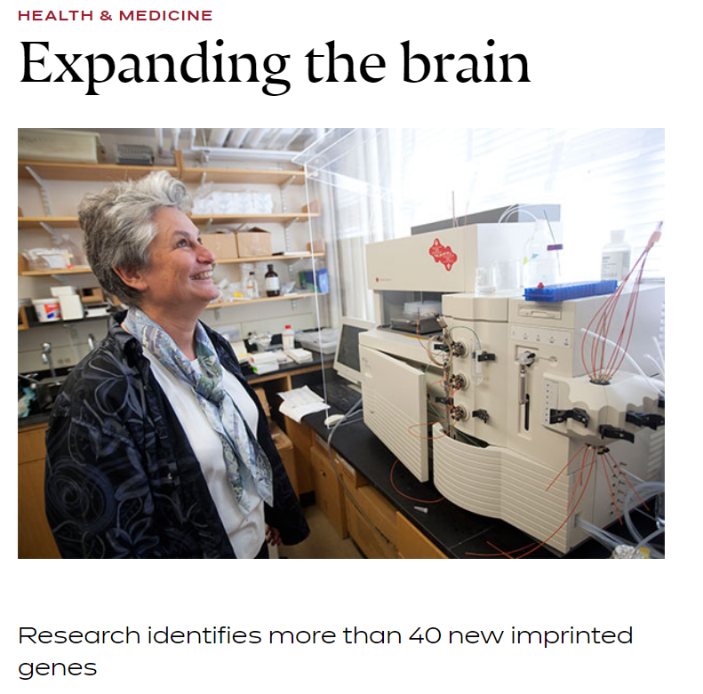
Research identifies more than 40 new imprinted genes.
Led by Catherine Dulac, the Higgins Professor of Molecular and Cellular Biology, a team of researchers has identified more than 40 new “imprinted’ genes, in which either the maternal or paternal copy of a gene is expressed while the other is silenced. (Full The Harvard Gazette article) File photo by Kris Snibbe/Harvard Staff Photographer

Aired May 25th. Features Catherine Dulac of our center in addition to Eric Kandel, Susanne Shultz, Charles Nelson, Margaret Spinelli and David Levine.

Story by Kate Yandell, May 13th. Discusses neuronal positioning system (NPS) recently developed by Jeff Lichtman's lab, published April 21 in Nature Methods.

Story by Stephanie Dutchen, May 21st. New method for visualizing chromosomes in astonishing detail, developed by Ting Wu's lab in collaboration with Xiawei Zhuang and Peng Yin's groups, published May 12 in Nature Communications.
More Stories
The Boston Children’s Hospital’s science and clinical innovation blog Vector (February 2019 by Nancy Fliesler), features the new paper titled “NMDA 2A receptors in parvalbumin cells mediate sex-specific rapid ketamine response on cortical activity”.
Read the Vector blog entitled “How the antidepressant ketamine rapidly awakens the brain and why its effects vary more in women”
Geographical maps have long changed the way we live, from the ancient hand-drawn ones that relied on explorer voyages to those on today’s satellite-driven, real time apps like Google Maps. The same transformative power holds true for maps in neuroscience. From the meticulous illustrations of neurons in different brain regions made by neuroanatomist Ramon y Cajal in the late 1800s to the imaging of neural circuits through more modern computerized technologies, the maps that we have of our nervous systems are constantly shaping the questions we think to ask and are able to answer about how our brains and bodies work—and what to do when diseases or injuries arise.
A recent advance in the map-making world of brain scientists comes from the lab of Xiaowei Zhuang, a faculty member in the Conte Center at Harvard who is the David B. Arnold Professor of Science and a Howard Hughes Medical Institute Investigator. In a study published in Cell, led by postdoctoral fellows Yaron Sigal and Colenso Speer, her team showcases a powerful new approach for visualizing and analyzing the input fields of neurons, using neurons in the eye called retinal ganglion cells as a test case. While mapping the sea of inputs that a single neuron receives and integrates is something that neuroscientists have long recognized as critical for understanding the functions of different kinds of neurons and their role in circuits—and have been able to accomplish in varying capacities—until now there’s usually been a trade-off between obtaining very high resolution images and having precise molecular information. The Zhuang team’s new strategy will allow researchers automated analysis of neuron input fields in a way that minimizes this compromise and overlays complex molecular information upon three dimensional reconstructions of neurons situated in relatively thick sections of brain tissue.

When it comes to science, perhaps the wisest amongst us are those who know how much we don’t know. The famous 19th century neuroanatomist Santiago Ramón y Cajal, who many would argue did more to characterize the different cell types that make up the nervous system than any other scientist of his time, once described the adult brain as a ‘jungle’ that is ‘impenetrable and indefinable.’
Over a hundred years later, despite an explosion of new technologies in microscopy and genetic and immunological methods of brain cell labeling, neuroscience research remains chock full of uncharted territories. In a recent publication in Cell, a team led by Jeff Lichtman, the Jeremy R. Knowles Professor of Molecular and Cellular Biology at Harvard—fittingly also named the Santiago Ramón y Cajal Professor of Arts and Sciences—presents the latest chapter in this story, by providing unparalleled views into the intricacies of synaptic connectivity in the cerebral cortex and revealing novel insights about this connectivity—while simultaneously highlighting just how much we have left to discover.
Original Research Article in eLife:
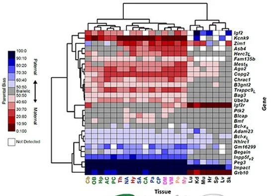
Quantitative and functional interrogation of parent-of-origin allelic expression biases in the brain.
By Perez, Rubinstein et al. July 3, 2015.
Harvard Gazette: Expanding the Brain. By Peter Reuell, July 29, 2015.

A new review explains how different methods of tissue clearing work
Imaging biological tissues is always a challenge because they are three dimensional, and issues of light scatter make it hard to clearly view deeper structures without cutting sections. Neuroscientists have long been interested in methods for “clearing” - or making more transparent -brain tissue so it is easier to image intact.
“I think as a neuroscientist there’s no part of the body where clearing is potentially more influential in moving forward,” says Jeff Lichtman, a professor of molecular and cellular biology and Conte lab head who teaches microscopy at Harvard and the Marine Biological Laboratory in Woods Hole.
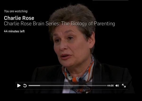
Aired May 25th 2015. Features Catherine Dulac of our center in addition to Eric Kandel, Susanne Shultz, Charles Nelson, Margaret Spinelli and David Levine.
Aired June 16th 2015. Features Catherine Dulac of our center in addition to Eric Kandel, Ben Barres, Norman Spack, Melissa Hines and Janet Hyde.

Harvard Medical School News: FISHing For Insight >>
Story by Stephanie Dutchen, May 21st. New method for visualizing chromosomes in astonishing detail, developed by Ting Wu's lab in collaboration with Xiawei Zhuang and Peng Yin's groups, published May 12 in Nature Communications.

BBC World Service:The Forum – Plasticity >>
Aired May 19th. Features Takao Hensch of our center talking about brain plasticity alongside an artist who works with plastics from garbage and a nanochemist who considers plasticity at the level of atoms and electrons. Host Bridget Kendall.
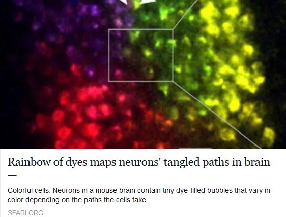
SFARI Autism News: Rainbow Of Dyes Maps Neurons’ Tangled Paths In Brain >>
Story by Kate Yandell, May 13th. Discusses neuronal positioning system (NPS) recently developed by Jeff Lichtman's lab, published April 21 in Nature Methods.
Circadian rhythms outside the brain’s central pacemaker may control the trajectory of brain plasticity
Scientists have long known that humans and other animals have a master biological clock situated in a deep brain region called the hypothalamus. In this master clock, cells manufacture timekeeper molecules whose levels oscillate to maintain the 24 hour cycles that define our lives. Yet these same timekeeper molecules are made throughout the brain and body, suggesting undiscovered functions for circadian genes outside the hypothalamus.

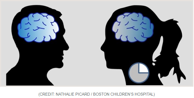
Sex-based differences in sensory processing identified at cellular level for first time
Take a moment to picture the reaction of a man in a bar to a beautiful woman walking in the door. Then picture the reaction of a woman sitting in the same spot in the same bar, looking at the same thing. Chances are you imagined different behaviors.
“The most extreme behavioral differences within a species are found in the way male vs. female animals respond to the same sensory stimuli,” write the authors of a recent study on sensory processing in the mouse brain, led by Catherine Dulac, Higgins Professor of Molecular and Cellular Biology in the Department of Molecular and Cellular Biology at Harvard and a Howard Hughes Medical Institute Investigator.
Neuroscientist Jeff Lichtman on Being Humbled by Nature
From drops of pond water to slivers of brain tissue, Jeff Lichtman has always enjoyed looking inside things. His interest in the microscopic realm began back when he was a young boy growing up in Westchester, New York. The son of a hematologist, he had a small research grade microscope in his bedroom from second grade on, complete with its own oil immersion lens.
“I kind of took it for granted. It didn’t even occur to me that this was bizarre,” he says.
Many people with autism display atypical sensory processing, and in independent studies, abnormalities in a deep brain structure known as the insula. A new study from researchers at Harvard reveals that across four different mouse models of autism, the development of sound-touch integration—important for social play in rodents—is impaired. This impairment reflects deficits in inhibitory circuit maturation in the insula and can be rescued by boosting inhibitory neurotransmission early in brain development.

DULAC TEAM IDENTIFIES HYPOTHALAMIC NEURONS GOVERNING PARENTAL BEHAVIOR IN MICE
Original journal article in Nature, “Galanin neurons in the medial preoptic area govern parental behaviour”
Boston Globe story, “Brain Circuits May Control Parental Behavior”
Harvard Gazette story, “Parental Controls”
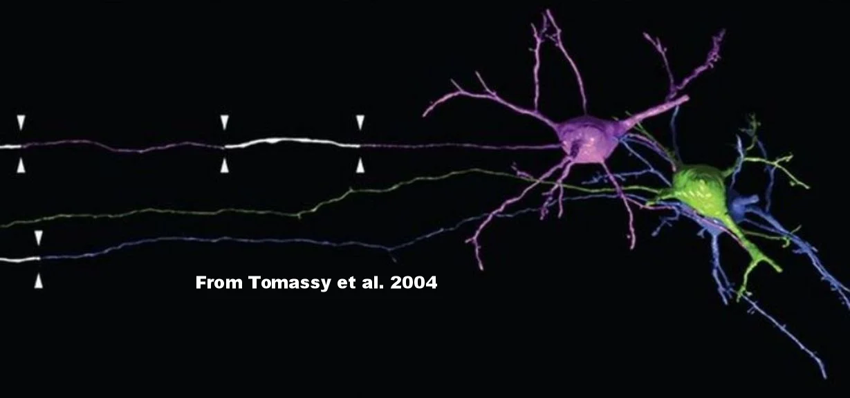
In collaborative study with Arlotta lab, Lichtman team characterizes myelin distribution of single cortical axons
Original Journal Article in Science:
“Distinct profiles of myelin distribution along single axons of pyramidal neurons in the neocortex“
Harvard Gazette story:
“Turning science on its head”

Listen to the interview on WBUR’s “On Point” Show with Tom Ashbrook:
“A Pill for ‘Brain Youth’?”
Listen to the interview on NPR’s Weekend Edition with Linda Wertheimer:
“Want Perfect Pitch? You Might Be Able To Pop A Pill For That”
Read the article in Frontiers in Systems Neuroscience that sparked this coverage:
“Valproate reopens critical-period learning of absolute pitch”
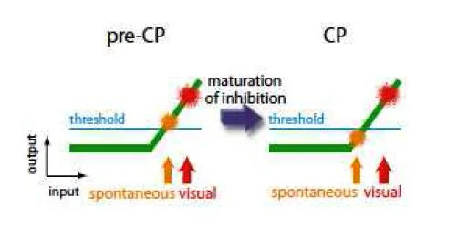
Model of visual critical period suggests that maturation of inhibition in early postnatal development lowers the ratio of spontaneous to evoked cortical activity
Inhibitory circuits in the cerebral cortex are known to mature subsequent to excitatory circuits, and this later maturation of inhibition is believed to play a central role in triggering critical periods. Yet exactly how the rise of inhibition “turns on” a critical period has long remained enigmatic.
In a Viewpoint article published October 2nd in Neuron, researchers propose that the answer lies in the selective suppression of spontaneous, as opposed to environmentally-evoked, neural activity. Critical period plasticity may be induced when there is a shift from predominantly internal to predominantly external learning cues, and the ratio of spontaneous to evoked cortical activity is lowered past a certain threshold…

Novel histone variant enforces ‘use it or lose it’ policy in olfactory neurons
Our experiences shape our identities. It matters if we grew up on a farm or in the city. If we’ve endured poverty or had material comforts. Even whether we’re exposed more to Disney films or horror movies, hip-hop versus classical music. Experiences both big and small personalize our nervous systems beyond what’s written in our DNA, and can lead to lasting physical changes in the brain. London cab drivers, for instance, after years of studying the intricate layout of streets in their city, develop larger hippocampi—the seahorse-shaped inner areas of our temporal lobes, important for memory and spatial navigation. But exactly how our nervous systems record and respond to our complex environmental experiences is still one of the biggest questions in neuroscience…

From discovery to trial: A drug that may correct ‘lazy eye’
A blog post by Sarah Lewin in Vector, Boston Children’s Hospital’s science and clinical innovation blog

A small group of highly metabolically active neurons act as conductors in the symphony that is cortical development. The movements of this symphony are the ‘critical periods’ of plasticity in which our experiences sculpt neural circuits—timed differently for different cortical areas and functions, but building upon each other in a precise sequence. The conductor neurons, known as parvalbumin cells (PV-cells), are a subset of inhibitory GABA interneurons that synchronize the activities of large populations of projection neurons across the cortex and direct the onset and closure of critical periods.

The skeletons of neurons have long fascinated scientists. Beyond giving these cells their unique shapes and structures, the cytoskeleton plays an active role in many processes that define neurons—from the birth of axons and dendrites to the development of synaptic sites and the dynamics of neurotransmitter release.

A throat culture can tell a doctor if someone has strep throat. A blood test if they have anemia. An X-ray if they have a broken bone. Problems in brain function, by contrast, are much harder to evaluate objectively.
Aired May 19th. Features Takao Hensch of our center talking about brain plasticity alongside an artist who works with plastics from garbage and a nanochemist who considers plasticity at the level of atoms and electrons. Host Bridget Kendall.